Don't miss our weekly Masters newsletter | Sign up now Don't miss our weekly Masters newsletter | Sign up now

  Medical Imaging - MSc/PGDip/PGCert


University of Leeds    School of Medicine

Full time & Part time September PGDip, MSc, PGCert 1 year full time, 2 years part time (MSc), 9 months full time, 2 years part time (PGDip), 9 months full time, 9 months part time (PGCert)
Engineering (12) Medicine (26)

FindAMasters summary

Are you passionate about medical imaging and its vital role in diagnosing and treating diseases? Look no further than our Medical Imaging programme at Leeds. Whether you're a recent graduate, a medical student wanting to intercalate, or a healthcare professional, this MSc/PGDip/PGCert course will equip you with the knowledge and skills needed for a successful career in the field. Tailor your studies to your interests with optional modules in MRI, Ultrasound, X-ray, CT, and more. Benefit from our renowned teaching and research expertise, and learn from world-leading academics. Join us on campus or online and unlock exciting career opportunities in medical imaging and related disciplines.

About the course

Medical imaging plays an essential role in modern medicine, supporting the diagnosis, treatment and monitoring of disease. At Leeds, we will build your understanding of the physics involved in different imaging techniques and enhance your imaging knowledge in preparation for your career.

Whether you are a recent graduate, a medical student wishing to intercalate, or a healthcare professional, our course will introduce you to:

Read more about this course

Entry Requirements

A bachelor degree with a 2:2 (hons) in a biological/physical science or radiography, or medicine or dentistry equivalent.

This course is also available as an intercalated programme to students who have completed three years of a UK medical degree and are normally ranked in the top 50% of their year of study. Read more at Intercalation.

The part-time variant of this programme is not suitable for international applicants who require a student visa. International applicants who do not require a student visa may be able to access the part-time variant of this programme by special arrangement.


Fees

UK: £12,750 (Total)
International: £31,000 (Total)

 Course Content

Visit institution website

Where will I study?

Where is University of Leeds

Videos


Student Profiles

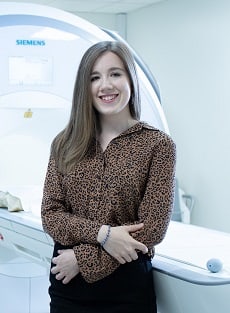
Rebecca Thornley


I am starting a job as a trainee medical physicist at Leeds Teaching Hospitals NHS Trust in September. The knowledge I obtained from this course helped me greatly in the interview process for this scheme.

I chose to study after studying physics at undergraduate level I became interested in the medical applications of physics so was looking for a course that would allow me to explore this. I liked the variety of modules offered by the course at Leeds, they covered a wide range of topics to suit a variety of backgrounds. Some modules e.g. forensic imaging offered an insight into an area of medical imaging I had never had the opportunity to learn about before.

I enjoyed being taught by lots of different academics and professionals who could offer perspectives on a topic eg academics, medical physicists, radiologists, radiographer etc. This made the modules really interesting and engaging. I also enjoyed studying alongside people of different background eg dental students, medical students, radiographers etc as everybody had their own unique insight into the field.

The highlights of the course have been the excellent teaching across modules and the support received from the module leads. I’ve also really enjoyed the opportunities to see the imaging equipment in action eg visiting the MRI department at Leeds General Infirmary and using ultrasound scanners on phantoms.

My project is titled ‘Multi-parametric MRI for radiotherapy planning and treatment evaluation in glioblastoma’. It has involved the analysis of MRI data acquired from 6 patients pre-treatment (post-surgery), during radiotherapy treatment (after 10 fractions) and six weeks post-treatment. This data includes T1 and T2 measures, diffusion and dynamic contrast-enhanced images in addition to conventional MRI data (T1-weighted, T2-weighted and FLAIR. I have been analysing the dataset using commercial and local software in order to assess the response of Glioblastoma to radiotherapy.

Doing a research project will benefit me in the future as I will have the opportunity to get involved in similar research projects during my job as a trainee medical physicist, so I will be able to apply the research skills I have obtained from this course directly to my job.

The medical imaging course at Leeds is well structured and provides a background to all the basic imaging modalities as well as giving you the opportunity to look into more specialised techniques or areas sp

Genevieve Liew

I have been working as a radiographer in Tan Tock Seng Hospital, Singapore for the past five years. Clinical work has been very fulfilling, and I was looking for a well-designed and comprehensive postgraduate programme to enhance my knowledge in medical physics. With its wide selection of modules, all of which were very relevant to radiography, the MSc in Medical Imaging offered by the University of Leeds seemed like the perfect choice. With a vibrant student community, the campus feels warm and inviting, with many communal areas to interact and study together with friends. Leeds on the whole is a very liveable city, with a walkable town centre, and great cafes and parks to hang out in.

I really enjoyed the holistic teaching approach of the programme, where we engaged with various academics, physicists, radiologists and radiographers. These different clinical perspectives made learning a very enriching experience. I particularly found the cross-sectional imaging modules: MRI, CT and Radionuclide Imaging very useful, with abstract concepts explained with depth and clarity, and the practical applications covered in detail. MATLAB visualisation sessions in X-ray served as great learning aids, while the Medical Interpretation module covered crucial imaging principles.

Readjusting to student life after having been working for the past few years, with multiple assignment deadlines and exams to prepare for was challenging in the beginning. We were all assigned personal tutors, who were very accommodating and helpful, meeting with us regularly to offer guidance and support.

The library facilities are great, with in-house cafes, numerous private study rooms and booths, and larger areas designated for group discussions. In particular, the Brotherton library has amazing architecture, with its own museum and art gallery – studying there can be quite atmospheric. The library website has a wealth of information on academic writing, referencing, and provides access to numerous journals and databases.

This programme has equipped me with a wealth of knowledge about the physics concepts underpinning the clinical modalities, and has helped develop my critical-thinking and research skills. I would highly recommend this course to any radiographer looking to gain a holistic understanding of radiography, and the various machines and scanners we use on a daily basis. I feel well-equipped with the knowledge gained from this course to better handle

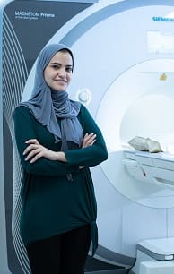
Bashair Alhummiany

I choose to study at the University of Leeds because of their reputation of excellence in the medical imaging field and high ranking among the universities in the UK.

Throughout the course I enjoyed learning from expert instructors that have worked in the fields for so many years, so they were successfully able to provide actual examples that illustrate the course concepts.

During the medical imaging courses, I developed my academic and research skills through completing the core modules. I have updated my knowledge on the advanced image analysis and post-processing techniques.

Perhaps the greatest challenge was studying with other students in this course who had different experiences and disciples. However, this diversity of students was actually very beneficial on my educational level, since it offered multiple perspectives in discussions.

My project involved preclinical PET-CT images of mice with abdominal aortic aneurysm disease using two segmentation techniques to estimate the Standardised uptake value (SUV) when a threshold value is applied.

Through working on a research project, I have established software development skills for data manipulation and modelling. I also had the chance to work as part of a research group through working closely with my supervisors, which had helped me in developing my skills and solve challenges.

It was interesting to work on different simulation software that had an impact on understanding the theory behind the practice. This is especially the case with the physics aspects of the modules, in order to aid a deeper understanding of the principles that cannot be gained from experience only. Also, many services provided by the university library helped boost my academic skills.

Following the MSc course, I am pursuing my interest in the field of medical imaging through doing a PhD. The international PhD Academy for cardiovascular and metabolic disease fascinated me and I am sure it will be a motivating atmosphere that will further develop my skills. It is amazing how the Academy provide a bespoke PhD programme that is particularly made to meet the required needs for a postgraduate researcher.

My PhD project involves analysing MRI images of the kidneys for patients with diabetic kidney disease to extract biomarkers that are hypothesised to detect changes in the kidney parenchyma at an early stage in order to improve the prediction of disease progression.

Upon co

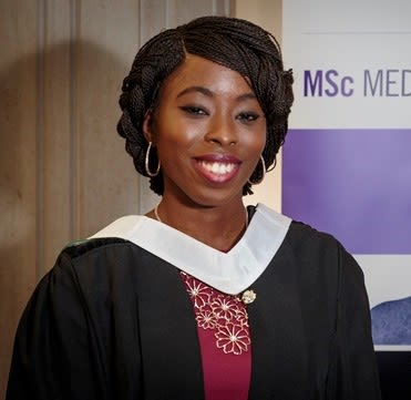
Ruth Abigobu-Alafun

I am from Nigeria and graduated from the University of Ghana with a BSc in Diagnostic Radiography in 2013 and I worked as a radiographer before my admission into the University of Leeds. I decided to study Medical Imaging at Leeds because of the structure of the course and the time frame.

The thing I enjoyed the most studying at the University of Leeds is the ease and comfort provided for studying, ranging from books to study spaces and the trips organised by LUU.

During my course, I learnt the following challenging topics; Application of Convolution and Filter, Quantification and Imaging Registration, Spin-Echo and Gradient Echo Imaging and Scan Parameters.

All the modules I studied were compulsory and interesting but Medical Image Analysis, MRI and Radionuclide Imaging stood out for me. These modules were not just new to me but also challenging and inspiring. They acted as an eye opener not only to current practices in the field but also the changes and development that are expected to happen in the future.

As a Radiographer, I feel Medical imaging provides a blend between the clinical aspect of imaging as well as a bit of engineering and in turn leading to a much better understanding and appreciation of image quality. It has also opened my mind to rapid development in imaging and how I can be involved as well as move with this fast growing field.

The University of Leeds also has top notch teaching facilities available for students. I found the library to be most influential in my study. It wasn’t as effective studying in my room as compared to the library. I recall staying back after lectures to either study or to do assignments in the Health Science Library. The closing time for the library was 12 am making it possible to take breaks and also read into the night which was an absolutely fantastic time for me to study.

The lecturers were very supportive. They were very approachable and very generous with their time. They were there to give further explanations or guidance through scheduled visits or emails. Now I have graduated I intend to implement what I have learnt as much as possible and also using this qualification as a ladder into furthering my career path in terms of obtaining a PhD.

Search Suggestions

Based on your current searches we recommend the following search filters